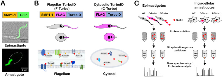
FIG 1.
T. cruzi life cycle and schematic of TurboID-expressing lines generated for proximity-dependent biotinylation experiments. (A) Live confocal images of SMP1-1–GFP localized to the flagellum of T. cruzi epimastigotes and an intracellular amastigote; the white oval denotes the position of the amastigote body. (B) Strategy for generating stable T. cruzi lines expressing TurboID in the flagellum using SMP1-1 as the endogenous bait protein or in the cytoplasm of epimastigotes and amastigotes, where addition of exogenous biotin mediates the biotinylation (red star) of proteins in close proximity to TurboID in both settings. The FLAG epitope is included to facilitate TurboID localization in transfectants. (C) Flow chart outlining the experimental protocol used for identification of biotinylated proteins in epimastigotes (left) and intracellular amastigotes (right). For both life stages, wild-type (WT), cytoplasmic-TurboID (labeled “C”), and flagellar-TurboID (labeled “F”) parasites (left to right) were exposed to biotin, and the biotinylated protein fractions in protein lysates were captured on streptavidin-agarose beads and subjected to mass spectrometry for identification and subsequent proteomic analysis.

