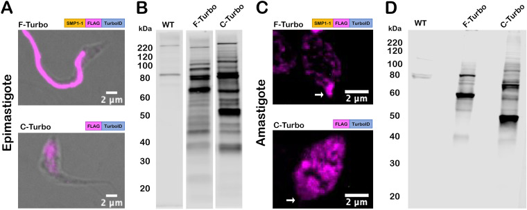
FIG 2.
TurboID localization and activity in T. cruzi. (A and C) Fluorescence microscopy images of fixed T. cruzi epimastigotes (A) or amastigotes (C) expressing SMP1-1-FLAG-TurboID (F-Turbo) (top) or FLAG-TurboID (C-Turbo) (bottom) stained for FLAG epitope (anti-FLAG) (pink). In panel C, white arrows indicate the position of the amastigote flagellum. (B and D) Biotinylated proteins in lysates of WT, F-Turbo, and C-Turbo T. cruzi epimastigote (B) and amastigotes (D) detected with streptavidin-DyLight 800.

