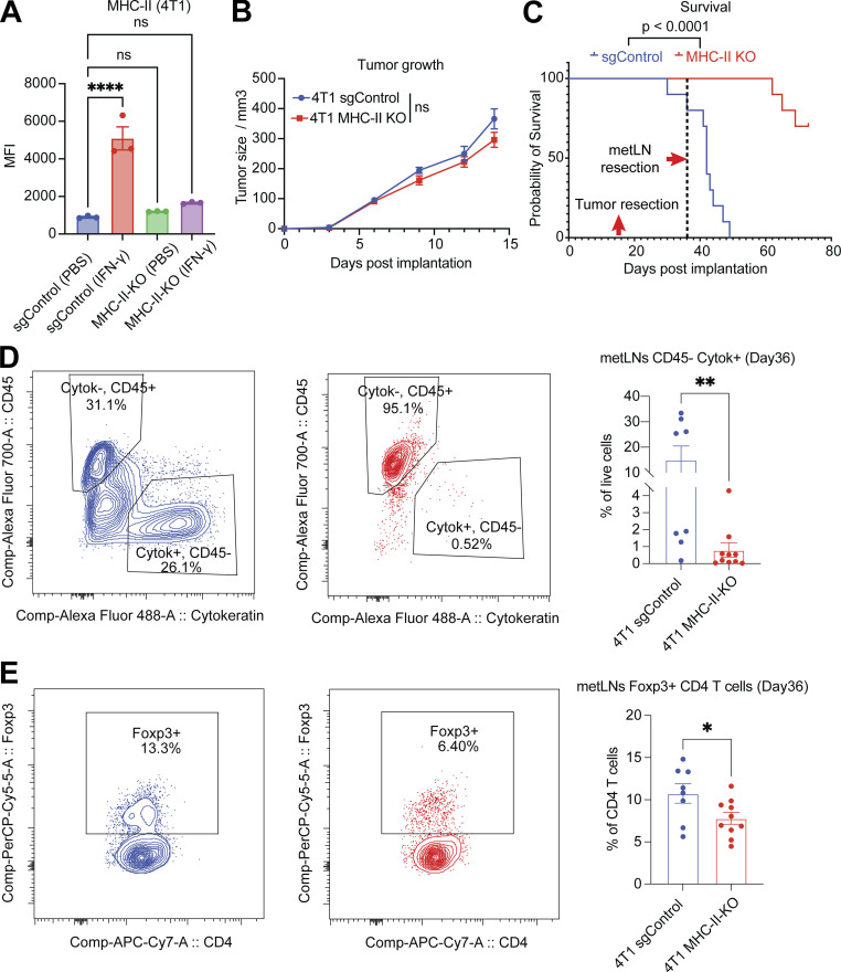
Figure 5.
The loss of MHC-II in cancer cells diminishes LNM. (A) The mean fluorescence intensity of cell surface MHC-II molecules in 4T1 sgControl and MHC-II knockout cells, with or without IFN-γ (10 ng/ml) treatment for 24 h, measured by flow cytometry. One-way ANOVA was used for statistical analysis, and Tukey’s multiple comparisons test was used for the post-hoc test. (B) The tumor growth of 4T1 sgControl (n = 10) and MHC-II knockout cells (n =10). Student’s t test was used for the statistical analysis. (C) Knockout of MHC-II in cancer cells increases the overall survival of mice. The primary tumors were removed on day 15, and the draining lymph nodes were removed on day 36. (D) Knockout of MHC-II in cancer cells led to a significant decrease in the proportion of cancer cells in the TDLNs. (E) Knockout of MHC-II in cancer cells resulted in a significant reduction in the proportion of Treg cells in the TDLNs. Student’s t test was used for statistical analysis; *, P < 0.05; **, P < 0.01; ****, P < 0.0001.

