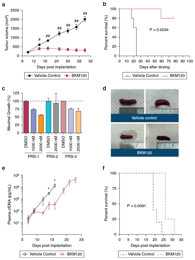
Figure 4. Antitumor activity of BKM120 in cell line and PDX xenografts.

(a, b) Effect of BKM120 on tumor growth and survival in HH xenografts (n = 5 per group). *P < 0.01, **P < 0.001 by unpaired t-test with one-tailed P-values. (c) GI of BKM120 in primary tumor cells from PDXs. (d–f) Comparison of spleen sizes and tumor burden measured by tumor cfDNA and survival in PRS-1 treated with BKM120 versus vehicle control (n = 5 per group). Tumor volume and cfDNA are presented as mean ± SEM and analyzed by unpaired t-test with one-tailed P-value (*P < 0.05). The survival curves were generated by the Kaplan–Meier method and analyzed by the Gehan–Breslow–Wilcoxon test. cfDNA, cell-free DNA; GI, growth inhibition; PDX, patient-derived xenograft.
