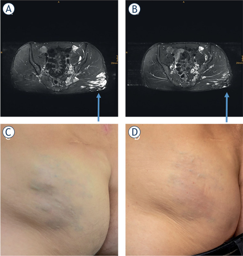
FIGURE 2.

Patient treated by BEST. Axial T2-weighted, fat-saturated MRI with hyperintense (arrow) gluteal venous malformation before treatment (A). Axial T2-weighted, fat-saturated MRI of the same region 4 months after treatment. The main part of the venous malformation is occluded (B). Photography before (C) and after the treatment (D).
