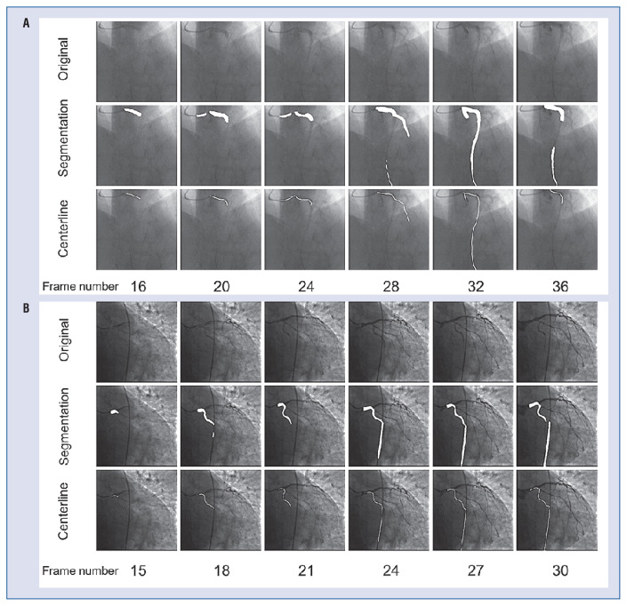
Figure 5.
Paradigmatic cases of unsuccessful automatic coronary flow reserve computations. The first row of each group is the original image, the second row is the segmentation result, and the third row is the extracted vessel centerline. Common mechanisms for failure are poor visualization of contrast dye (A), and mis-segmentation of the catheter (B) or of other anatomic structures.

