
Figure 3.
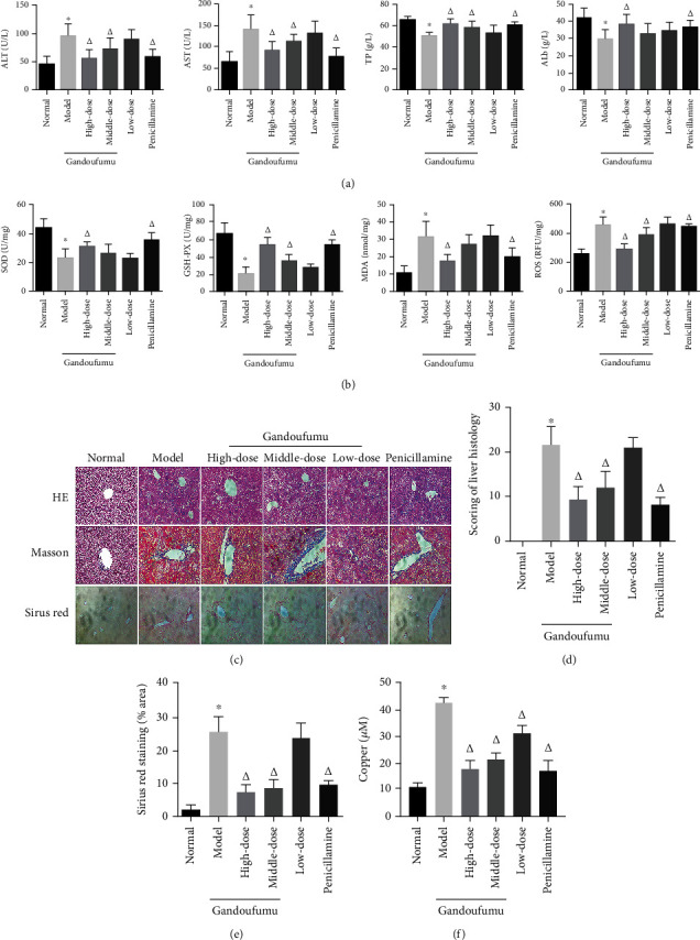
Effect of GDFMD on serum liver function, liver oxidative stress, pathological liver changes, and serum copper content of mouse models of WD. Serum levels of ALT, AST, TP, and ALB (a); liver oxidative stress of SOD, GSH-Px, MDA, and ROS was measured by ELISA assays (b). Representative photomicrographs of morphological changes (c) in HE (400x), Masson (400x), and Sirius red (400x) staining in liver tissues, scoring of liver histology (d), and quantification of Sirius red staining area (e) of mice from the control, model, Gandoufumu (high dose, low dose, and middle dose), and penicillamine groups. Serum copper content was determined by copper assay kit (f). Data is depicted in terms of mean ± SD (n = 10). ∗P < 0.05 compared with the normal group; △P < 0.05 compared with the model group.