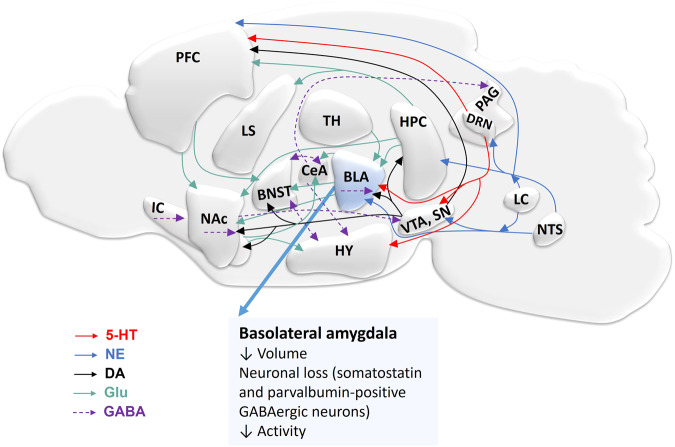
Fig. 1. Overview of neuronal circuits involved in fear and anxiety in the rodent brain.
The basolateral amygdala (BLA) is a central regulator of fear circuitry and other brain regions are involved in these processes. Interconnection of various brain regions in fear by different neurotransmitter pathways including serotonergic (red), glutamatergic (green), dopaminergic (black), norepinephrinergic (blue), and GABAergic (purple). Efferent projection of serotonin from dorsal raphe nucleus (DRN) to various brain regions such as BLA, ventral tegmental area (VTA), substantia nigra (SN), and prefrontal cortex (PFC). Glutamate projections interconnect different brain regions. Norepinephrine (NE) is released from projections of locus coeruleus (LC) and nucleus tractus solitarius (NTS). The VTA and SN provide dopaminergic inputs to the nucleus accumbens (NAc), bed nucleus of the stria terminalis (BNST), BLA, hippocampus (HPC), PFC, and insular cortex (IC). VTA and SN receive GABAergic projections from NAc and the central amygdala (CeA) sends GABAergic projections to the hypothalamus (HY) and the periaqueductal gray (PAG). BNST sends or receives GABAergic projection to/from HY and CeA; and GABAergic interneurons are present in NAc, IC, and BLA. Reduced amygdala volume, neuronal cell loss, and neuronal activity are associated with anxiety disorders. Serotonin: 5-hydroxytryptamine (5-HT); gamma-aminobutyric acid (GABA); dopamine (DA); glutamate (Glu); lateral septum (LS); thalamus (TH).

