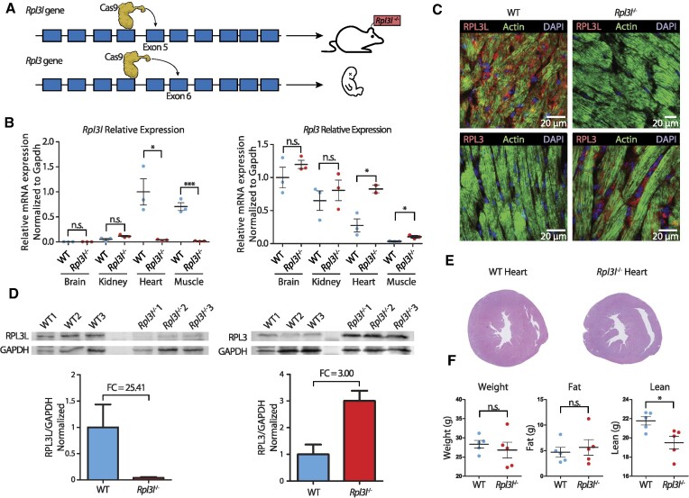
Figure 2.
Phenotypic and molecular characterization of Rpl3l−/− knockout mice. (A) Strategy for generation of Rpl3−/− and Rpl3l−/− mice using the CRISPR/Cas9 system. Rpl3l−/− mice were successfully generated by introducing a 13 bp deletion in exon 5. Rpl3−/− mice have an embryonic-lethal phenotype. See also Supplementary Figure S1. (B) Relative expression levels of Rpl3l (left) and Rpl3 (right) measured using RT–qPCR and normalized to Gapdh. Rpl3 is ubiquitously expressed, while Rpl3l is heart and muscle specific. Rpl3l−/− mice do not express Rpl3l in any of the tissues (n = 3). Statistical significance was assessed using the unpaired t-test (*P <0.05, **P <0.01, ***P <0.001). (C) Immunofluorescence staining of RPL3 and RPL3L in both WT (left) and Rpl3l−/− mice heart tissues (right). Nuclei have been stained with DAPI and are shown in blue; actin is depicted in green and RPL3L (uL3L) (top) and RPL3 (uL3) (bottom) in red. (D) Western blot analysis of RPL3L (uL3L) (left) and RPL3 (uL3) (right) in cardiomyocytes isolated from WT and Rpl3l−/− hearts (n = 3 and n = 6, respectively). At the bottom, barplots depicting the fold change of RPL3 (uL3) and RPL3L (uL3L) expression in cardiomyocytes is shown. RPL3L (uL3) and RPL3 (uL3) levels were normalized to GAPDH. See also Supplementary Figure S4 for full blot images, and Supplementary Figure S5 for western blot results using total heart samples from WT and Rpl3l−/− mice. (E) Representative histological sections of WT and Rpl3l−/− heart tissues stained with H&E. A total of 10 mice were included in the histological analyses. See also Supplementary Figure S9. (F) EchoMRI analyses of aged (55-week-old) WT and Rpl3l−/− mice, in which weight, fat and lean mass were measured for each animal (n = 5). Statistical significance was assessed using unpaired t-test (*P <0.05).

