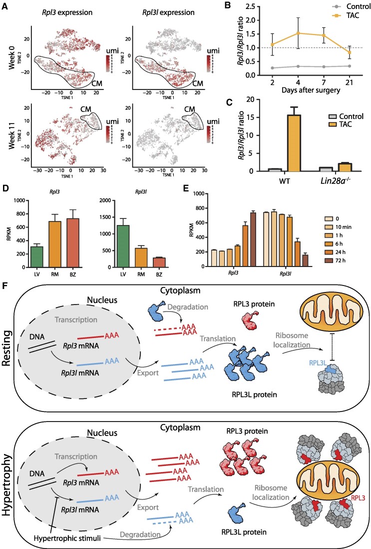
Figure 6.
Pressure overload leads to an increase in Rpl3 expression and a decrease in Rpl3l expression in the heart. (A) The expression of Rpl3 and Rpl3l across cell types in mouse hearts before (week 0) and after hypertrophy (week 8) induced by transverse aortic constriction (TAC). Cardiomyocytes (CM) are circled. Expression levels are shown as umi (unique molecular identifiers). (B) TAC leads to heart hypertrophy that correlates with increased Rpl3 expression and decreased Rpl3l expression. The Rpl3/Rpl3l ratio is significantly increased at all time points (2, 4, 7 and 21 days after surgery) when compared with control hearts. See also Supplementary Figures S19 and S20 and Supplementary Table S15. (C) Effects of TAC-induced hypertrophy on Rpl3–Rpl3l interplay are impaired in Lin28a−/− mice. See also Supplementary Figure S22. (D) Rpl3 (left) and Rpl3l (right) nuclear mRNA levels in cardiomyocytes from the left ventricle (LV), remote myocardium (RM) and border zone (BZ) after myocardial infarction. Processed data (RPKM) were obtained from Günthel et al. (81). (E) Rpl3 and Rpl3l mRNA expression levels at different time points (0 min, 10 min, 1 h, 6 h, 24 h and 72 h) after myocardial infarction. Processed data (RPKM) were obtained from Liu et al. (82). (F) Model showing the Rpl3–Rpl3l interplay in resting (top) and hypertrophic (bottom) conditions. In the resting heart, Rpl3l is predominantly expressed in cardiomyocytes, and the RPL3L protein negatively regulates Rpl3 expression, while RPL3L (uL3L)-containing ribosomes do not establish close contact with mitochondria. Upon hypertrophic stimuli, Rpl3l expression is impaired and Rpl3l mRNA is degraded in the cytoplasm, leading to an increased expression of Rpl3. RPL3 (uL3)-containing ribosomes establish close contact with mitochondria.

