Figure 3.
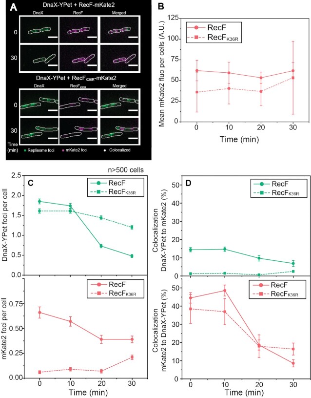
RecF-mKate2 over-expression increases replisome dissociation. The effect of RecF-mKate2 over-expression on replisome stability was determined by two-color single cell imaging. Strains used are deleted the recF gene, expressed a fluorescently tagged version of the clamp loader (DnaX-YPet), and carried a vector encoding the mKate2 tagged versions of RecF. Cells were loaded into home-built flow chamber incubated at 37 ºC and imaged as described in the method section. (A) Colocalization imaging between mKate2 (RecF or RecFK36R) and the replisome (DnaX-YPet). Images were obtained in the single channels 568 nm (mKate2) and 514 nm (DnaX-YPet), then merged. Imaging of single cells before and 30 min after arabinose addition. (B) Evolution of the mKate2 fluorescence signal per cell over the 30 min of over-expression. The values represented are the mean ± SEM, at time 0, 10, 20 and 30 min after induction, with n > 500 cells for each strain. (C) Number of replisome and mKate2 (RecF) foci detected during the 30 min following the over-expression. (D) Colocalization percentage during the 30 min of over-expression of one fluorophore foci relative to the other and vice versa. The values represented are the mean value ± SEM.

