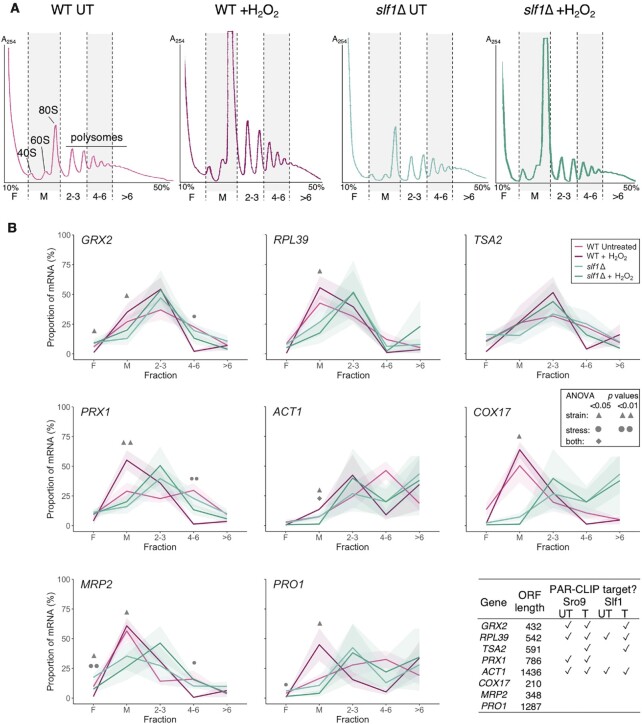
Figure 4.
mRNAs remain ribosome associated during stress in slf1Δ cells. (A) Polysome profiles from 15–50% sucrose gradient fractionated cell extracts with stripes indicating five pooled fractions collected for qRT-PCR: F = ribosome free, M = monosome 2–3/4–6/>6 = increasing polysome association. Traces coloured as per key in panel B. (B) Proportion (%) of individual mRNAs found in each polysome fraction by qRT-PCR in WT (magenta) or slf1Δ (cyan) in optimal growth conditions or following 15 min H2O2 treatment (darker shades). The shaded uncertainty envelope around each line represents the s.e.m. (n = 3). A two-way ANOVA with a Tukey post hoc test was used to assess the effect of the strain and the stress and to test the interaction of these two factors for each gradient fraction. Symbols (defined in the inset key) indicate significant results. Table shows which RNAs are PAR-CLIP targets in each dataset (✓).

