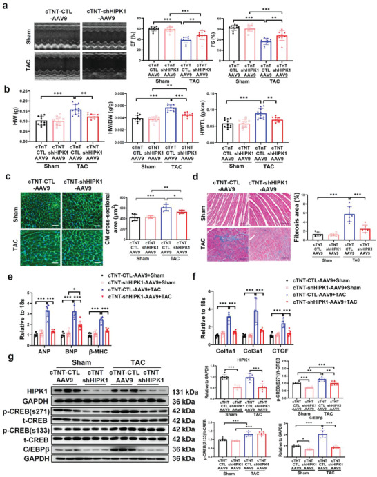
Figure 5.

cTnT‐AAV9‐mediated HIPK1 knockdown prevents TAC‐induced pathological cardiac hypertrophy. a) Echocardiography for LV ejection fraction (EF) and fractional shortening (FS) at 6 weeks after TAC surgery (n = 10–11). b) Heart weight (HW), heart weight/body weight (HW/BW) ratio, and heart weight/tibia length (HW/TL) ratio of mice (n = 10–11). c) Wheat germ agglutinin (WGA) staining for cardiomyocyte (CM) cross‐sectional area (n = 6). Scale bar = 50 µm. d) Masson staining for cardiac fibrosis area (n = 6–7). Scale bar = 100 µm. e,f) qRT‐PCR for e) ANP, BNP, and β‐MHC and f) Col1a1, Col3a1, and CTGF in mice heart tissues after sham or TAC surgery (n = 6). g) Western blot for HIPK1, CREB phosphorylation, and C/EBPβ expressions in mice heart tissues with sham or TAC surgery (n = 6). Data among four groups were compared by two‐way ANOVA test followed by Tukey post hoc test. *, p < 0.05; **, p < 0.01; ***, p < 0.001.
