
Figure 3.
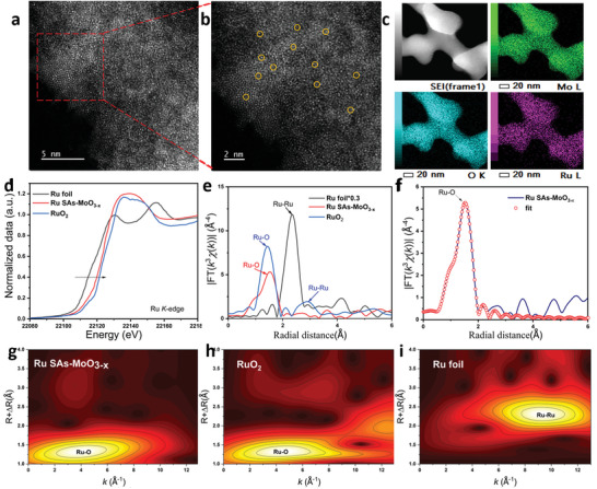
Structure characterization of Ru single atom in Ru SAs‐MoO3− x . a,b) HAADF‐STEM images of Ru SAs‐MoO3− x . c) HAADF‐STEM image and related elemental mapping images of Ru SAs‐MoO3− x . d,e) Ru K‐edge XANES and EXAFS for Ru SAs‐MoO3− x , RuO2, and Ru foil. f) R‐space fitting curve for Ru SAs‐MoO3− x . g–i) WT for the k2‐weighted EXAFS signal for Ru SAs‐MoO3− x , RuO2, and Ru foil.