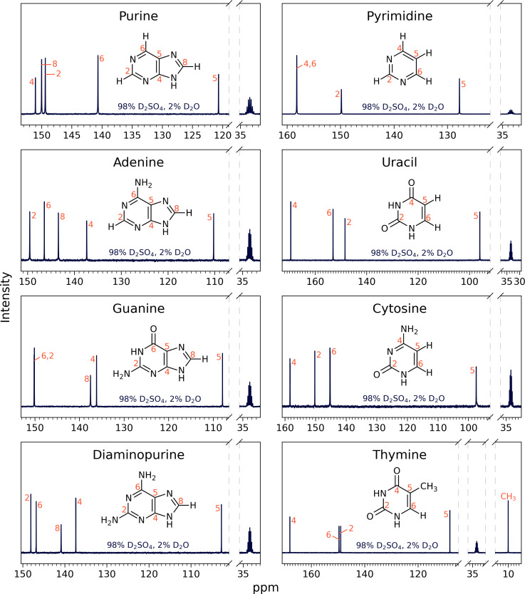
Fig. 3.
13C NMR spectra for eight nucleic acid bases: purine, adenine, guanine, diaminopurine, pyrimidine, uracil, cytosine, and thymine in 98% D2SO4/2% D2O (by weight) with DMSO-d6 as a reference, at room temperature. The labeled NMR carbon peaks match the number of carbon atoms in the known molecular structure for a given compound. All peaks are consistent with the molecules being stable and the structure not being affected by the concentrated sulfuric acid solvent. For a description of peak assignments, see Section 2.3., Figs. 5 and 6, and SI Appendix, Figs. S1–S10.

