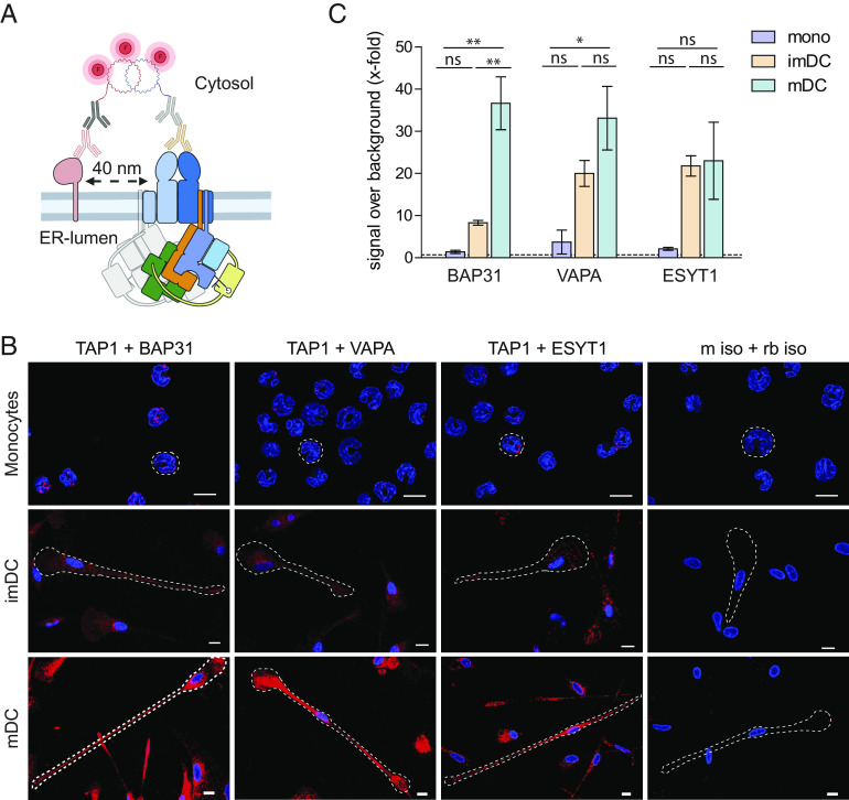
Fig. 4.
BAP31, VAPA, and ESYT1 are in 40 nm proximity to TAP in imDCs and mDCs. (A) Schematic representation of proximity ligation assay (Duolink). Briefly, target proteins are immunolabeled with primary antibody raised in two different species; then, incubation with secondary antibodies coupled to an oligonucleotide sequence “PLUS” or “MINUS” follows. If target proteins are in 40 nm proximity, complementary oligo sequences are ligated, and the polymerase proceeds to a concatemeric amplification of the DNA template finalized with the hybridization of fluorescently labeled oligos to the amplified sequence. (B) For microscopy, moDCs were chemically fixed with 3% (v/v) formaldehyde/PBS, and a proximity ligation assay was performed with TAP1 (mAb 148.3) and BAP31, VAPA, or ESYT1. As control, corresponding isotype antibodies were used. Nuclei were stained with DAPI for visualization (blue). (Scale bar, 10 μm.) (C) For flow cytometry, moDCs were fixed and semipermeabilized, and a proximity ligation assay was performed with TAP1 (mAb 148.3) and BAP31, VAPA, or ESYT1. Median fluorescence intensity increase ± SD was plotted as x-fold over background (dash line). Two donors per experimental setup were analyzed. Statistical analysis was performed using one-way ANOVA with Turkey post hoc. *P ≤ 0.05 and **P ≤ 0.005, ns: not significant.

