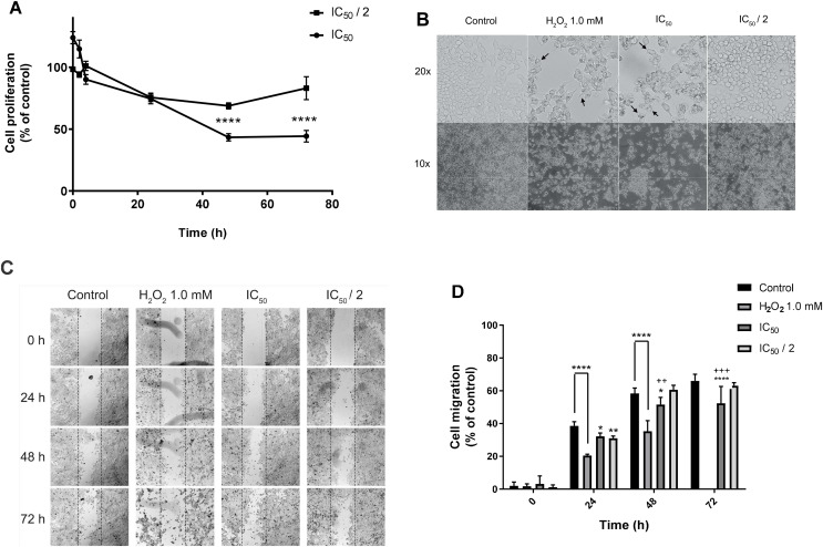
Figure 3. Effect on cell proliferation, morphology, and migration.
MCF7 human breast cancer cells were treated with IC50 and IC50/2 values of 7-hydroxy-3,4-dihydrocadalene for 48 h, and then: (A) Trypan blue assay was performed to measure cell proliferation and (B) cell imaging multi-mode reader Cytation 5 was used to study cell morphology. (C) Wound healing in MCF7 monolayer at 0, 24, 48, and 72 h after treatment (10×). The dotted lines indicate the edge of the initial wound. (D) Cell migration separation area was quantified at different times with each treatment and compared with the percentage of reduction of the initial area (0 h). The results were expressed as a percentage of migration capacity of the cells with respect to time zero of each group. The results are shown as means ± SD of at least three independent experiments (*P < 0.05, **P < 0.01, ****P < 0.0001 vs. Control and ++P < 0.01, +++P < 0.001 vs. IC50/2).

