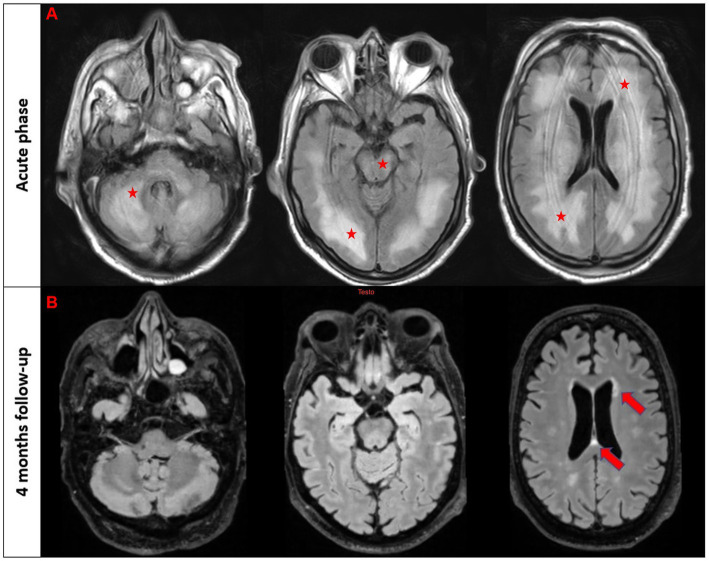
Figure 1.
T2/FLAIR imaging in the acute phase (first raw) and follow-up brain MRI after 4 months in patient #2. The first three images from the acute phase reveal a diffuse T2/FLAIR hyperintensity involving bilateral white matter, brainstem, and cerebellum (marked with stars). The follow-up scans demonstrate a nearly complete resolution of these abnormalities with few scattered white matter hyperintensities in the periventricular regions (marked with an arrow).

