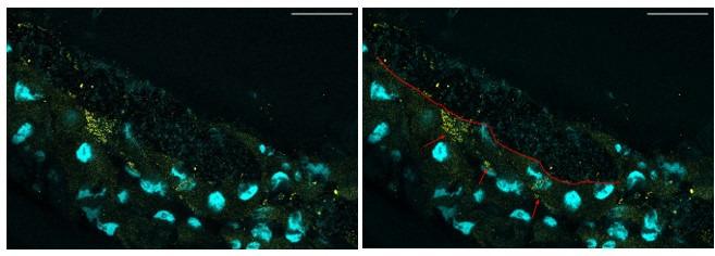
Author response image 2. Representative confocal picture of the anterior midgut of a Lp-associated larva.

We selected a plane showing putative inclusions of Lp’s rRNA inside the enterocytes. Scale bar: 50 µm. Right picture: the red line depicts the border between the lumen (on the top, characterized by DAPI-positive bacteria) and the gut epithelium (on the bottom). The red arrows show putative inclusion of Lp’s 16S rRNA in the enterocytes.
