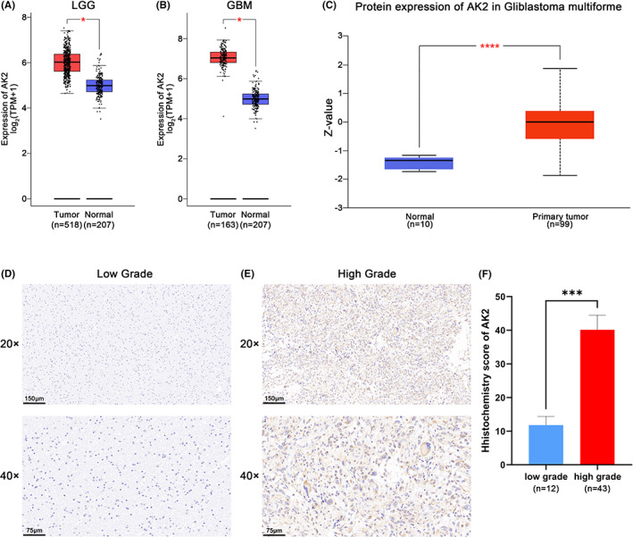
FIGURE 1.

AK2 Expression of mRNA and protein level between gliomas and normal brain tissues. (A) Different expression of AK2 at mRNA level between LGG and normal brain tissues in GEPIA2 website. (B) Different expression of AK2 at mRNA level between GBM and normal brain tissues in GEPIA2 website. (C) Different expression of AK2 at protein level between gliomas and normal brain tissues in CPTAC database. (D, E) AK2 immunohistochemical pictures of the low‐grade and high‐grade glioma samples. (F) Immunohistochemistry revealed higher expression patterns of AK2 in high‐grade glioma tissues than in low‐grade glioma tissues. (*p < 0.05, **p < 0.01, ***p < 0.001, ****p < 0.0001).
