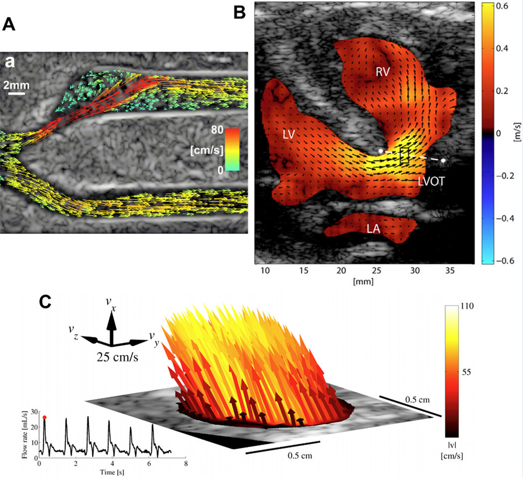
Figure 4.

(A) Vector projectile imaging (VPI) showing peak systole92. Copyright (2014), World Federation for Ultrasound in Medicine & Biology, reprinted with permission.
(B) Vector flow image of a patient (8 days) indicating a ventricular septal defect from a parasternal long-axis view99. Copyright (2014), World Federation for Ultrasound in Medicine & Biology, reprinted with permission.
(C) 3-D vector flow from the common carotid artery in peak-systole116. Copyright (2017), IEEE, reprinted with permission.
