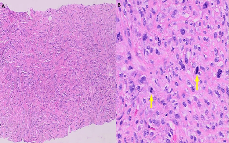
Figure 3. Ultrasound-guided core needle biopsy of left thigh mass displayed A) pleomorphic spindle cell sarcoma, high-grade with tumor cells arranged in storiform pattern (H&E, x100), B) on higher magnification sheets of polygonal, spindled, and epithelioid cells are seen, with eosinophilic cytoplasm, marked nuclear pleomorphic, multinucleation and conspicuous mitotic activity (yellow arrows), including atypical forms (H&E, x400).
H&E: hematoxylin and eosin stain

