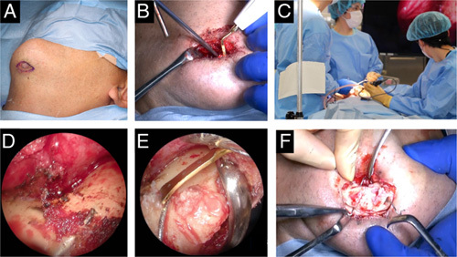
FIGURE 3.

Intraoperative views. (A) Fistula removal. (B) Excess bone removal using ultrasonic cutting instruments. (C) Situations using endoscopes. It is possible for surgeons and assistants to share a narrow surgical field of view. (D) Endoscopic image on the lingual side. The expanded field of view enables clear confirmation of the condition of bones and wires. (E) Surrounding bone cutting using an ultrasonic cutting instrument in an endoscopic view. (F) Labial wire removal was finally accomplished after lingual bone removal.
