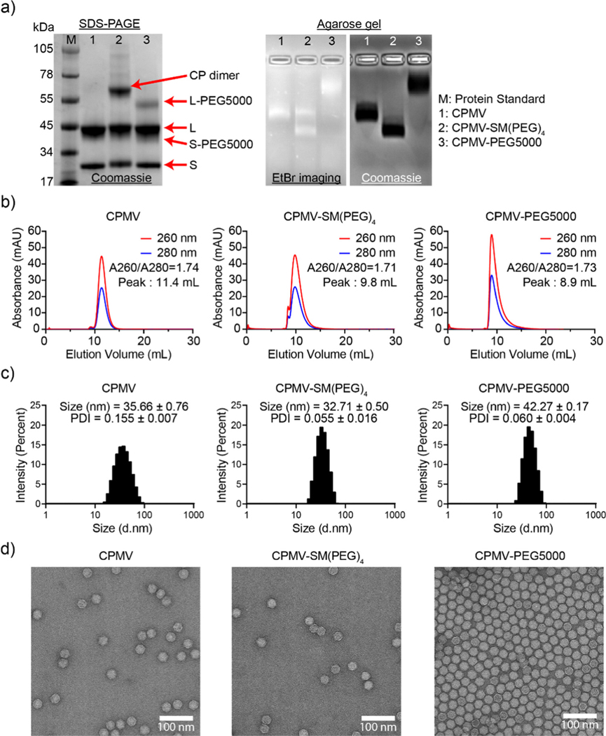
Fig. 2.
Characterization of CPMV and its bioconjugates. (a) SDS-PAGE (left) and agarose gel electrophoresis (right) of CPMV, CPMV-SM(PEG)4, and CPMV-PEG5000. SDS-PAGE shows the S (24 kDa) and L (42 kDa) proteins of CPMV. (b) SEC elution profiles. The inset shows the absorbance ratio of 260 nm/280 nm – a ratio of ~1.8 at the elution peak indicates intact CPMV. (c) DLS showed a single peak of CPMV, CPMV-SM(PEG)4, and CPMV-PEG5000, indicating a homogeneous particle distribution. The insets provide average sizes and polydispersity index (PDI) of three measurements. (d) TEM images of negatively stained (UF) CPMV, CPMV-SM(PEG)4, and CPMV-PEG5000 particles. Scale bar = 100 nm.

