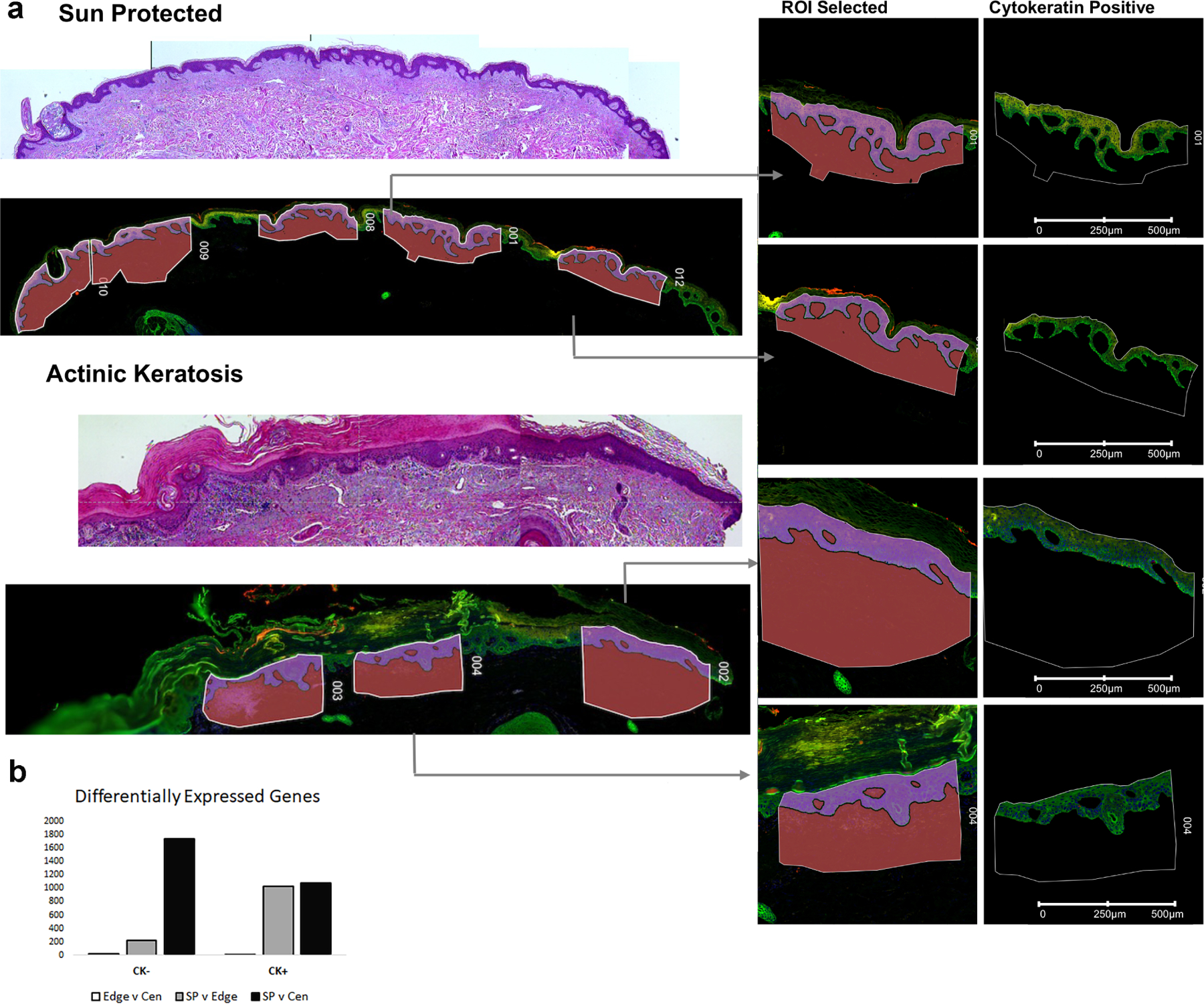
Figure 1: Spatial Profiling identifies the greatest transcriptional change in the cytokeratin negative compartment comparing sun-protected skin with actinic keratosis.

a: Digital Spatial Profiler identified regions of interest (ROI) highlights examples of areas of patient tissue sampled for transcriptional profiling. b: Differentially expressed genes identified using a pairwise FDR adjusted p-value of less than 0.05 and an overall t-test statistic of 0.05 highlights greater number DEGs (y-axis) in the cytokeratin negative compartment (CK-) comparing sun protected normal skin (SP) with actinic keratosis center (Cen). CK+ = cytokeratin positive compartment, Edge = actinic keratosis edge.
