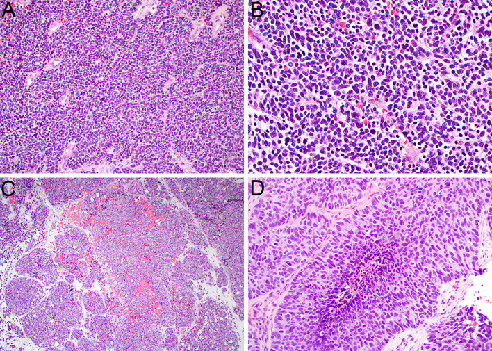
Fig. 2.
Sinonasal small cell neuroendocrine carcinoma displays sheets of markedly atypical epithelial cells with scant cytoplasm (A, × 20). The nuclei are hyperchromatic with frequent molding, mitotic figures, and apoptotic bodies (B, × 40). Large cell neuroendocrine carcinoma tends to show nested architecture (C, × 10). The tumor cells have abundant cytoplasm and large nuclei with vesicular chromatin and prominent nucleoli that demonstrate peripheral palisading (D, × 40)

