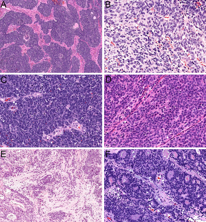
Fig. 3.
Olfactory carcinoma is predominantly composed of sheets and lobules of neuroectodermal cells (A, × 4). These cells have syncytial cytoplasm that ranges from scant to abundant (B, × 40). The nuclei are hyperchromatic and angulated with prominent nuclear molding and cell-cell wrapping (C, × 40). Most cases display overt neural differentiation with variable amounts of neurofibrillary matrix (D, × 40) including occasional expansile neurofibrillary zones with ganglion cells (E, × 10). Many cases also show well-formed true rosettes (F, × 20)

