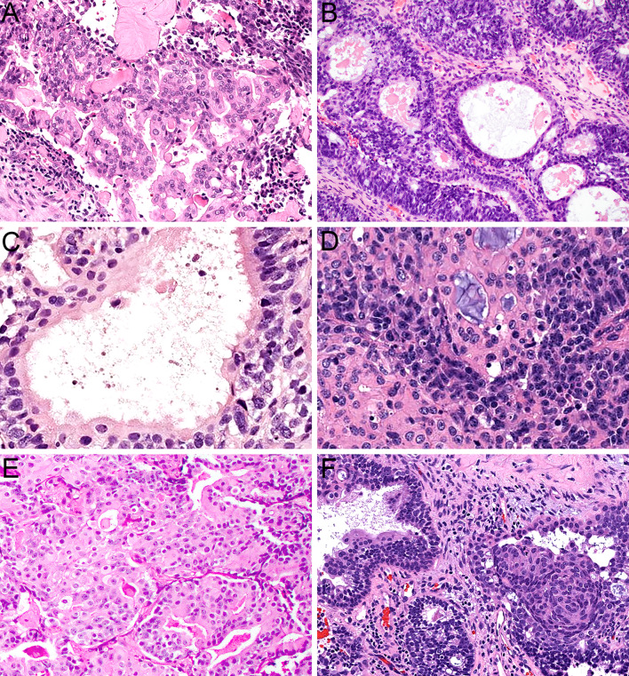
Fig. 4.
Olfactory carcinoma also displays a well-developed glandular component that can include either complex, expansile proliferations of back-to-back acini (A, × 20) or simple ducts and tubules that are closely intermixed with surrounding neuroectodermal cells (B, × 20). Many of the glands show well-developed cilia formation (C, × 40). The glandular cells have abundant eosinophilic cytoplasm and round to oval nuclei that lack the atypia of surrounding neuroectodermal cells (D, × 40). It can be difficult to differentiate glands from rosettes in some cases (E, × 20). Rare cases show squamous morules intermixed with the glands (F, × 20)

