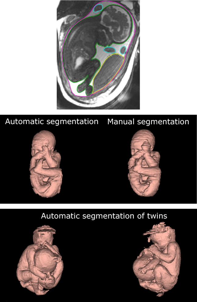
Fig. 1.

Segmentation of the fetus in magnetic resonance images. The top panel shows a magnetic resonance image with manual delineations of the fetus (green), placenta (yellow), umbilical cord (blue), and uterine wall (pink). This was repeated throughout the 3D image stack and all pixels in the image stack were classified as fetus, placenta, umbilical cord, or amniotic fluid. This pixel classification was used for training and evaluation of the proposed artificial neural network. The middle panel shows fetal 3D models generated by automatic (left) and manual (right) segmentation of the same fetus. The time required to generate the automatic model is 45 s, whereas the time required to generate an accurately manually segmented model is 1–2 h. Agreement between manual and automatic fetal segmentation is high (c.f. Fig. 3). The bottom panel shows the performance of the automatic method on twin fetuses. The proposed automatic fetal segmentation method was tested on a case of twin fetuses as proof of concept to show generalizability. Although the algorithm had only been trained on singleton fetuses, it shows promising generalizability. Artifacts at the top of one of the fetal heads are related to image artifacts in the 3D MRI images
