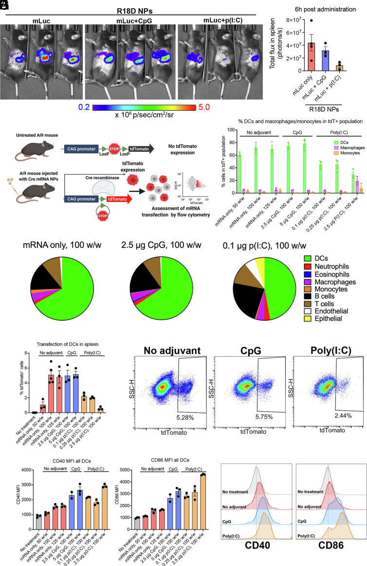
Fig. 5.
In vivo transfection in spleen following systemic administration of R18D mRNA nanoparticles (NPs). (A) R18D NPs carrying luciferase mRNA (mLuc) (10 μg/mouse) and CpG (2.5 μg/mouse) or poly(I:C) (0.1 μg/mouse) were assembled at a polymer-to-nucleic acid ratio of 100 w/w and administered intravenously to C57BL/6J mice. Whole-animal bioluminescence imaging was performed 6 h after administration. (B) Image analysis was used to assess total flux in the spleen. (C) Schematic of the Ai9 mouse model used to assess transfected cell types in vivo following systemic administration of mRNA NPs carrying Cre mRNA. Cells that are transfected undergo Cre recombinase–mediated recombination, resulting in tdTomato expression that is detected by flow cytometry. (D–H) R18D Cre mRNA NPs were administered intravenously to Ai9 mice at 10 μg mRNA/mouse, and tdTomato expression in key cell populations in the spleen was assessed after 24 h. (D) Percent of all tdTomato+ (tdT+) cells in the spleen that are DCs, macrophages, or monocytes. (E) Pie charts indicating average share of transfected cells in the spleen belonging to each cell population shown for NP treatments carrying no adjuvant, 2.5 μg CpG/mouse, or 0.1 μg poly(I:C)/mouse. (F) Percent of DCs in the spleen that are transfected. (G) Representative flow cytometry plots showing transfected tdTomato+ DCs treated with mRNA-NP formulations coencapsulating no adjuvant, 2.5 μg CpG, or 0.1 μg poly(I:C). (H) Geometric mean fluorescent intensity (MFI) of CD40 and CD86 expression in all splenic DCs. (I) Representative histograms of CD40 and CD86 expression in no-treatment control, and following NP treatment coencapsulating no adjuvant, 2.5 μg CpG/mouse, or 0.1 μg poly(I:C)/mouse. Error bars represent SEM.

