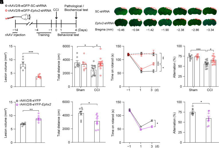
Fig. 2.
Manipulation of hepatic sEH using tail vein-virus injection modulated CCI-induced neurological deficits. (A) Schematic diagram of the experimental design. Black circles, control animals; red circles, adult C57BL/6J mice injected with rAAV2/8-eGFP-Ephx2-shRNA. (B–F) The effects of knocking-down hepatic Ephx2 following CCI. (B) Representative images of Nissl staining for brain injury. The red outline showing the loss area of injured brain. (C) Quantitative analysis of brain lesion volume (n = 6). (D) Spontaneous locomotor activity was assessed using the OFT (sham, n = 7; CCI, n = 14). (E) Sensorimotor function was assessed using the rotarod performance test at 1 d pre-CCI and 1, 3 d post-CCI (dashed line for sham group, n = 7; and solid line for CCI group, n = 14). (F) Spatial memory ability was tested using the Y-maze test (sham, n = 7; CCI, n = 14). (G–J) The effects of overexpression of Ephx2 in the liver in CCI. Black circles, control animals; purple circles, adult C57BL/6J mice injected with rAAV2/8-eYFP-Ephx2. (G) Quantitative analysis of brain lesion volume (n = 7). (H) OFT, (I) rotarod performance test, and (J) Y-maze test in rAAV2/8 virus-injected mice after CCI (n = 8). Statistical significance was determined using two-tailed unpaired Student’s t test for C, G, H, and J; two-way ANOVA followed by the Bonferroni’s post hoc-test for D and F; or two-way repeated measures ANOVA followed by the Bonferroni’s post hoc-test for E and I. Data are presented as the means ± SEM. Asterisks indicate significant differences (*P < 0.05, **P < 0.01, ***P < 0.001).

