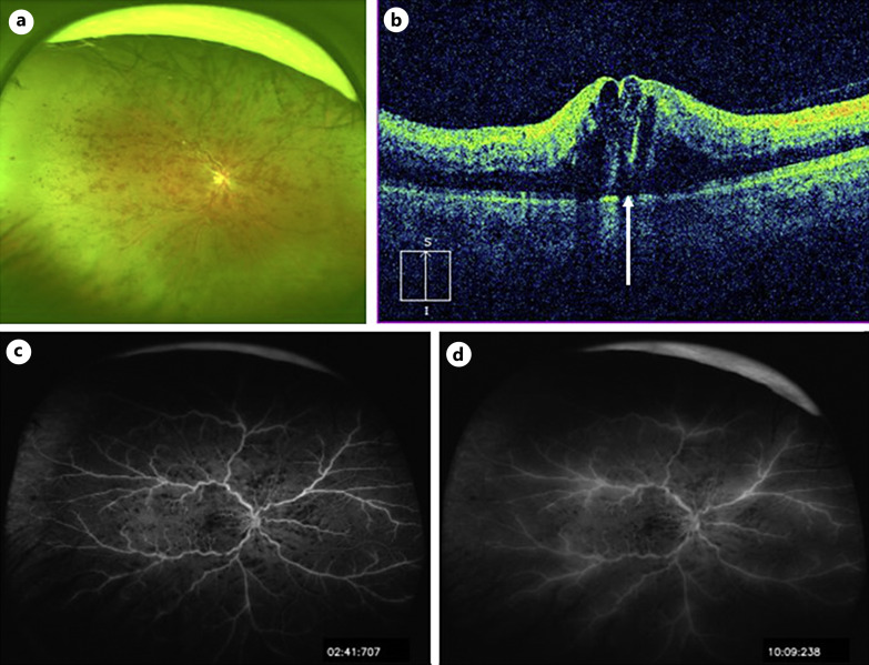
Fig. 1.
Baseline findings. a An ultra-widefield fundus photograph taken with Optos California (Nikon Corporation, Tokyo, Japan). Multiple retinal hemorrhages are observed throughout the fundus, and retinal veins are tortuous and dilated. b Vertical section of optical coherence tomography (OCT) image taken with Cirrus HD-OCT (Carl Zeiss Meditec, Dublin, CA, USA). Cystoid macular edema and small serous retinal detachment (arrow) are observed. c, d Ultra-widefield fluorescein angiograms taken with Optos California (c early phase, d late phase). Multiple hypofluorescent spots that appeared to be fluorescent block due to retinal hemorrhages (c, d) and hyperfluorescent leakage from the retinal veins are observed (d).

