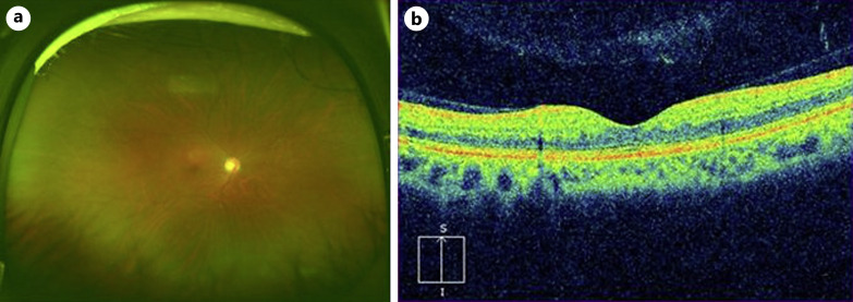
Fig. 3.
Findings 10 months after the onset. a An ultra-widefield fundus photograph taken with Optos California (Nikon Corporation, Tokyo, Japan). Retinal hemorrhages almost resolved. b Vertical section of optical coherence tomography (OCT) image taken with Cirrus HD-OCT (Carl Zeiss Meditec, Dublin, CA, USA). Cystoid macular edema and serous retinal detachment disappear.

