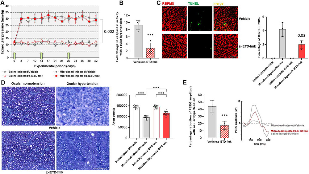
Fig. 1. Experimental modeling of ocular hypertension-induced glaucoma in rats and pharmacological inhibition of caspase-8 cleavage.
A. Intraocular pressure curves through the 6-weeks experimental period. Green arrows show the time points for z-IETD-fmk injections. Microbead injections resulted in a significant increase in intraocular pressure (P = 0.002) that did not change with z-IETD-fmk treatment or injections of vehicle only (P = 0.56; n > 40/group). B. Caspase-8 activity assay detected a significant decrease in z-IETD-fmk treated retinas (***P < 0.001; n > 4/group). C. TUNEL labeling (green) in retinal whole mounts showed a significant decrease (P = 0.03) in the number RBPMS-labeled RGCs (red) in z-IETD-fmk-injected ocular hypertensive eyes than vehicle-injected ocular hypertensive controls (n > 4/group; scale bar, 100 μm). D. Axon counts in optic nerve cross-sections indicated approximately 40% protection against ocular hypertension-induced axon loss with z-IETD-fmk treatment (***P < 0.001; n > 36/group; scale bar, 10 μm). E. The PERG amplitude was also preserved in z-IETD-fmk-treated ocular hypertensive eyes compared to vehicle-injected ocular hypertensive controls (n > 4/group). Data are presented as mean ± SD, P values were obtained using a one-way ANOVA.

