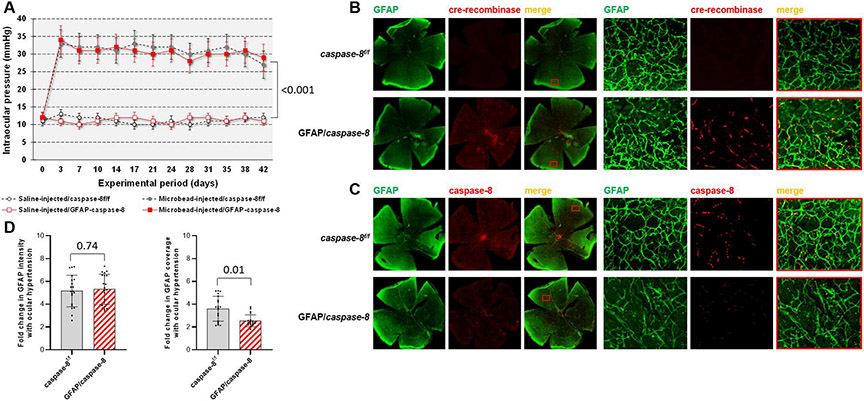
Fig. 3. Experimental modeling of ocular hypertension-induced glaucoma in mice and cre/lox-based deletion of astroglial caspase-8.
A. Intraocular pressure curves through the 6-weeks experimental period. Microbead injections resulted in a significant increase in intraocular pressure (***P < 0.001) that was similar in GFAP/caspase-8 mice and caspase-8f/f controls (P = 0.20; n > 39/group). B. Immunolabeling of retinal whole mounts demonstrated cre-recombinase (red) expression in GFAP+ astroglia (green) after tamoxifen-injection in GFAP/caspase-8 mice. C. Based on immunolabeling of retinal whole mounts, no caspase-8 (red) expression was detectable in GFAP+ astroglia (green) in GFAP/caspase-8 mice. Red boxed areas are shown in higher magnification (scale bar, 100 μm). D. Image analysis detected no alteration in the intensity of GFAP immunolabeling (reflecting the GFAP expression level of astroglia) with caspase-8 deletion. However, the coverage of GFAP immunolabeling (reflecting the size and density of individual cells) was significantly lower in GFAP/caspase-8 retinas compared with caspase-8f/f controls (n > 20/group). Data are presented as mean ± SD, P values (obtained using a one-way ANOVA) for the statistical comparison of experimental groups are given in each graph.

