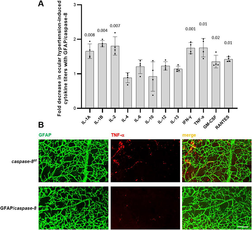
Fig. 7. Effects of GFAP/caspase-8 on pro-inflammatory cytokine production.
A. The bar graph presents the fold decrease in ocular hypertension-induced cytokine titers with GFAP/caspase-8 relative to caspase-8f/f (statistical significance are shown by P values obtained using a one-way ANOVA). Data (mean ± SD) were obtained using a minimum of three new samples per group. B. Immunolabeling of retinal whole mounts supported decreased cytokine response (TNF-α, red) of GFAP-labeled astroglia (green) to ocular hypertension in GFAP/caspase-8 mice compared to caspase-8f/f ocular hypertensive controls (scale bar, 100 μm).

