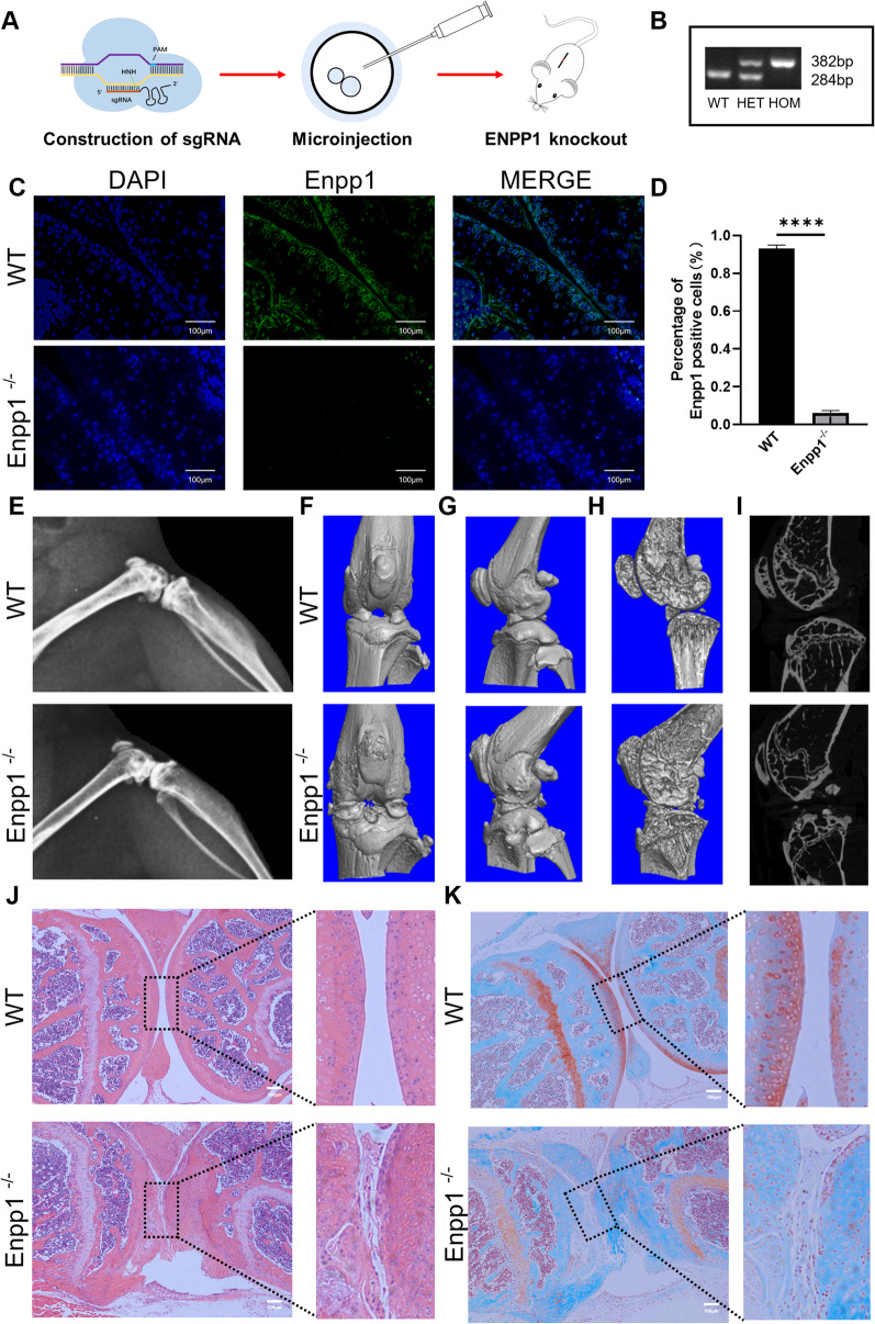
Fig. 1.
Enpp1 deletion caused knee OA: A Schematic for the construction of ENPP1 knockout (Enpp1−/−) mice using CRISPR/Cas9 technology. B The results of genotyping. C IF staining of Enpp1 in murine knee joint. (Enpp1, green; DAPI, blue; Scale bar: 100 µm, n ≥ 3). D The quantification of the expression of Enpp1 in WT and Enpp1−/− knee joint (WT groups = 93.10% ± 1.90%, Enpp1−/− groups = 6.03 ± 1.32, n = 3, p < 0.0001). E Representative lateral X-ray of WT and Enpp1−/− knee joints at week 14 (n ≥ 3). F–I Representative micro-CT scan of WT and Enpp1−/− knee joints at week 14. J H&E staining of WT and Enpp1−/− knee joint at week 14 (Scale bar: 100 µm, n ≥ 3). K Safranin O-Fast Green staining of WT and Enpp1−/− knee joints at week 14 (articular cartilage, red; bone, green; Scale bar: 100 µm, n ≥ 3).WT, wide type; HET: heterozygote; HOM homozygote; Enpp1, ectonucleotide pyrophosphatase/phosphodiesterase 1; IF, Immunofluorescence; DAPI, 4′,6-diamidino-2-phenylindole; WT, wild type; H&E, Hematoxylin & Eosin

