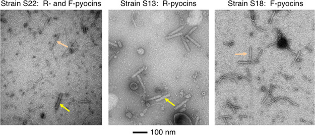
FIG 1.
TEM images of lysates of cells producing R- and F-type pyocins. A lysate of strain S22 (left panel), which produces both R- and F-type pyocins; a lysate of strain S13 (middle panel), which produces just R-type pyocins; and a lysate of strain S18 (right panel), which produces just F-type pyocins, are shown. R-type pyocin particles are indicated by yellow arrows, and F-type pyocin particles are indicated by orange arrows. Grids were negatively stained with uranyl acetate. The scale bar shown applies to all three micrographs.

