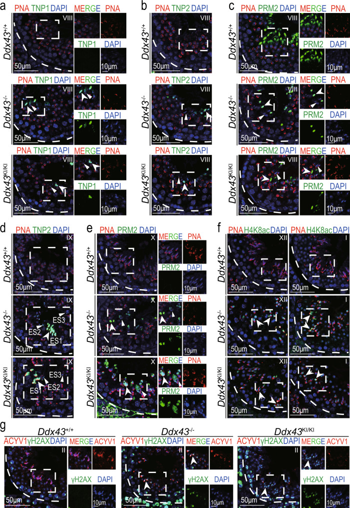
Fig. 3. DDX43 deficiency impairs chromatin remodeling during spermiogenesis.
a–g Immunofluorescence analyses on adult Ddx43+/+, Ddx43KI/KI and Ddx43–/– mouse testes with the following combinations. a TNP1 (green) and PNA (red), see also Supplementary Fig. 4a; (b, d) TNP2 (green) and PNA (red), see also Supplementary Fig. 4b; (c, e) PRM2 (green) and PNA (red); (f) H4K8ac (green) and PNA (red), see also Supplementary Fig. 5a, b; (g) γH2AX (green) and PNA (red). Right panels show monochromatic images of the boxed area in the left panels (a, c, e, g). Stages of seminiferous tubule are denoted by Roman numerals. White arrows indicate positive signals; White arrowheads indicate negative signals. DNA was counterstained with DAPI. Scale bars are indicated. A minimum of three animal samples were used for each genotype and each experiment was repeated three times with similar results.

