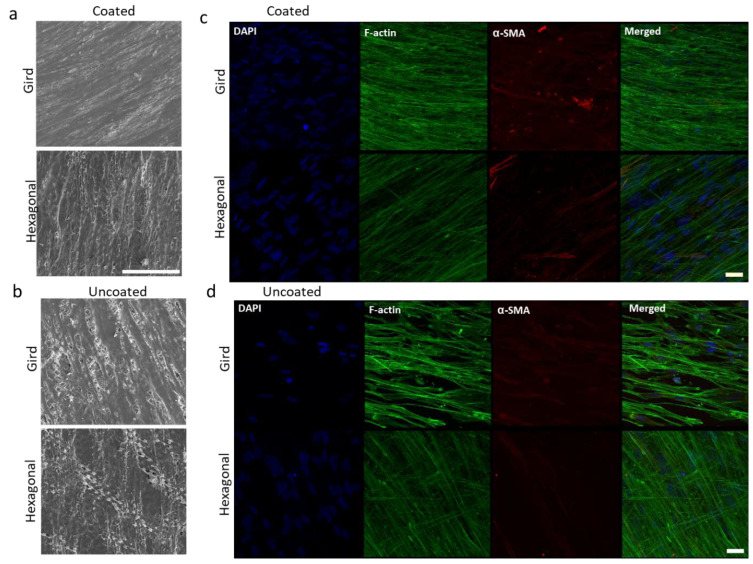
Figure 5.
Effects of gelatin coating on HTM cells grown on micropatterned porous PCL scaffolds for 14 days. (a,b) SEM images of HTM cells grown on PCL scaffolds. Scale bar = 100 µm. (c,d) Confocal imaging of F-actin (green) and α-SMA (red) expression of HTM cells grown on PCL scaffolds. Scale bar = 30 µm. (a,c) Coated with gelatin. (b,d) Uncoated controls. Blue: Nuclei.

