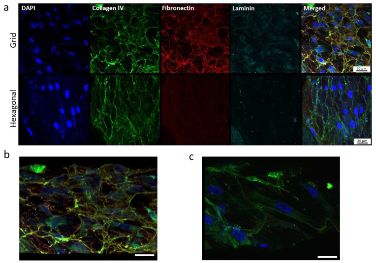
Figure 7.
Confocal images showing the effect of pore structures on ECM deposition. (a) Expression of ECM proteins, collagen, fibronectin and laminin of HTM cells grown on gelatin-coated, micropatterned, porous PCL scaffolds. (b,c) Tilted angle view of 3D reconstruction of HTM cells grown on the grid-patterned PCL scaffold (b) and hexagon-patterned PCL scaffold (c). Green, collagen IV; red, fibronectin; cyan, laminin; blue, DAPI-stained nuclei. Scale bar = 30 µm.

