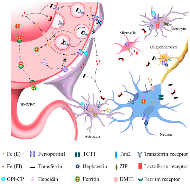
Figure 1.
Roles of different cells in brain iron metabolism. The main route of brain iron uptake is where the iron in the blood crosses the blood–brain barrier (BBB) via Tf-TfR1 in the apical surface of brain microvascular epithelial cells (BMVECs) and FPN1 in the basal surface of BMVECs. Iron can also enter the brain through the transcytosis of ferritin by its receptors at BBB. After iron influxes into the brain parenchymal tissue, it can enter astrocytes through their end feet surrounding BBB and then be transferred to neurons. Iron across the BBB can also directly enter the interstitial fluid of the brain and be transferred to neurons and other cells without passing through astrocytes (see black lines). Astrocytes hepcidin secreted through its end feet to directly decrease FPN1 level of BMVECs, which decreased the iron influx into brain tissues. GPI-CP expressed by astrocytes assists FPN1 in releasing iron into the brain. Astrocyte-specific Cp knockout blocks iron influx FPN1-CP pathway into the brain (see black lines and crosses). Neurons acquire both trivalent and divalent iron through TfR1, TCT1 and DMT1, while those astrocytes that are not part of the BBB acquire iron via DMT1 and ZIP molecules. Oligodendrocytes mainly uptake iron via DMT1 and Tim2. Oligodendrocytes can secrete Tf, while the activated microglia can secrete Lf. Neurons and glia store iron in ferritin and release iron through FPN1 with the coordination of CP/hephaestin or hepcidin, thereby further promoting cross-talk and interaction with other types of cells.

