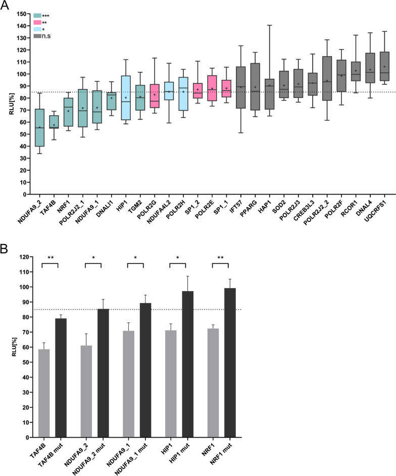
Fig. 2.
Automated dual luciferase reporter gene assays. A HEK 293T cells were transfected with 50 ng/well of either reporter plasmid pMIR-RNL-TK, with or without insert, and 200 ng/well of miRNA expression plasmid containing either the respective miRNA or no insert. The luciferase activities of the miR-34a transfected samples were normalized with respect to the luciferase activity measured with empty reporter plasmids. Four independent experiments were carried out in duplicates. Columns colored in turquois show a significant reduction of the luciferase activity with a p-value ≤ 0.001. Columns colored in magenta show a significant reduction of the luciferase activity with a p-value ≤ 0.01 and ≥ 0.001. Columns colored in violet show a significant reduction of the luciferase activity with a p-value ≤ 0.05. Columns colored in dark blue show a non-significant reduction of the luciferase activity with a p-value ≥ 0.05. Data are shown as mean ± SEM. B Dual luciferase reporter gene assays with mutated reporter plasmids. The criterion for a positive target gene was defined with a significant reduction (p-value ≤ 0.05) of the RLU of at least 15%. HEK 293 T cells were co-transfected with miR-34a-5p expression vectors and the wild type reporter plasmids of the respective target genes (light grey) or mutated reporter plasmids (mut) of the respective target genes (black) as depicted in the diagram. Four independent experiments were carried out in duplicates. Three asterisks represent a significant reduction of the luciferase activity with a p-value ≤ 0.001. Two asterisks represent a significant reduction of the luciferase activity with a p-value ≤ 0.01 and ≥ 0.001. One asterisk represents a significant reduction of the luciferase activity with a p-value ≤ 0.05. Ns indicates a non-significant reduction of the RLU

