Figure 8.
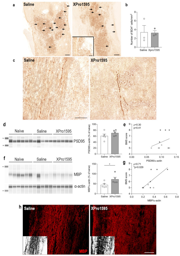
Selective inhibition of solTNF preserves myelin but does not alter the density of biotinylated dextran amine (BDA)+ cells and neuronal plasticity after SCI. (a) Representative images demonstrating corticospinal tract BDA+ axonal debris (arrows) engulfed by phagocytic-like cells located within the lesion area 24 days after SCI. (b) Estimation of the number of BDA+ cells/mm2 in the lesion area (n = 3/treatment group). (c) Representative images demonstrating BDA+ corticospinal axons with anterogradely transported BDA rostral to the lesion 24 days after SCI. (d) Quantification of post-synaptic PSD95 35 days after SCI. (e) Correlation analysis of PSD95 levels and motor function (BMS) 35 days after SCI. (f) Quantification of MBP 35 days after SCI. (g) Correlation analysis of MBP levels and motor function (BMS) 35 days after SCI. Protein data are normalized to α-actin expression and presented as a percentage of naïve mice (n = 6) with n = 5 mice/treatment group. White circles represent saline-treated mice and grey circles represent XPro1595-treated mice. * p < 0.05. (h) Representative images of MBP-labeled myelin structures in the peri-lesion area 35 days after SCI. Inserts show binary masks of MBP staining. Scale bars: (a,h) = 100 µm, and insert in (a,c) = 40 µm. Data are presented as mean ± SEM. MBP, myelin basic protein; PSD95, postsynaptic density protein 95; SCI, spinal cord injury.

