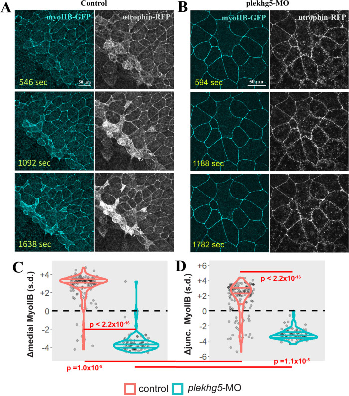
FIGURE 6:
Knockdown of plekhg5 prevents medial accumulation of MyoIIB. (A, B) Unlike cells in control embryos, in plekhg5 knockdown embryos, initial F-actin dynamics remains in the apical cortex but MyoIIB fails to show up in the medial domain. No actomyosin enrichment is observed with progression of time. (C, D) Quantification of medial and junctional MyoIIB intensity shows that unlike in control bottle cells where the intensity of MyoIIB increases, medial and junctional MyoIIB decrease in cells with plekhg5 knockdown. Each dot is an individual cell. Horizontal lines within each violin delineate quartiles along each distribution. s.d. = standard deviations. P values were calculated via a KS test. Data were collected from four control and five plekhg5 knockdown embryos.

