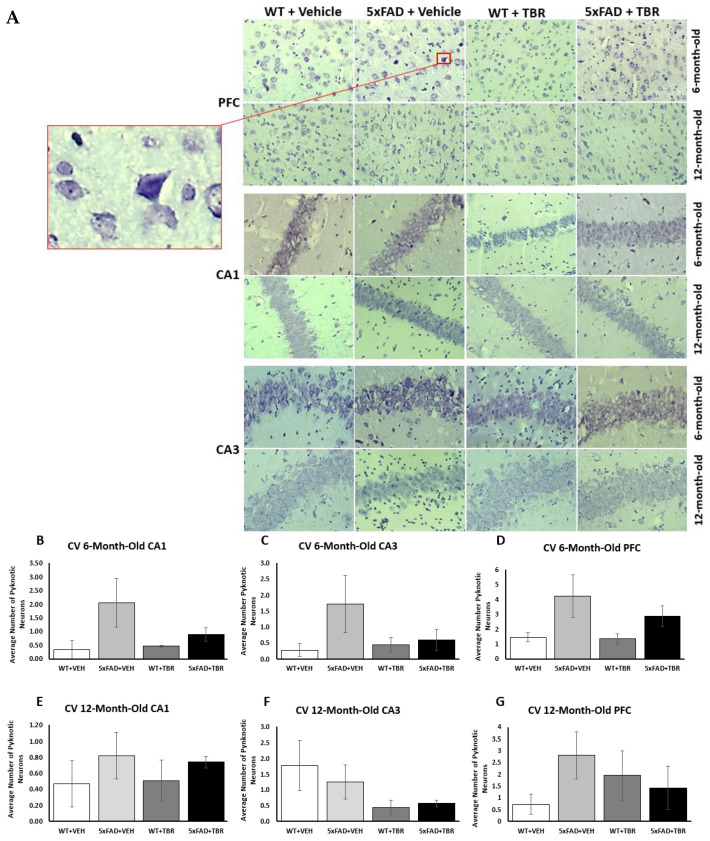
Figure 9.
Cresyl-violet stained tissue revealed pyknotic cells (see insert) in the pre-frontal cortex (PFC) and the CA1 and CA3 regions of the hippocampus in all mice (A). There was a non-significant trend toward more pyknotic cells in the 5xFAD mice in all but the CA3 area of the 12-month mice and treatments of TBR had no significant effects (B–G).

