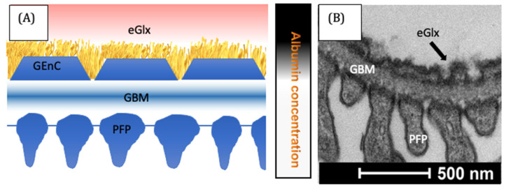
Figure 2.
The glomerular filtration barrier. In the representative diagram, (A) the filtrate passes the layers of the filter. Albumin is largely excluded, as indicated by the local albumin concentration on the right. Electron microscopy (B) demonstrates the functional arrangement of cells within the glomerulus [84]. GBM = glomerular basement membrane, GEnC = glomerular endothelial cells, PFP = podocyte foot process.

