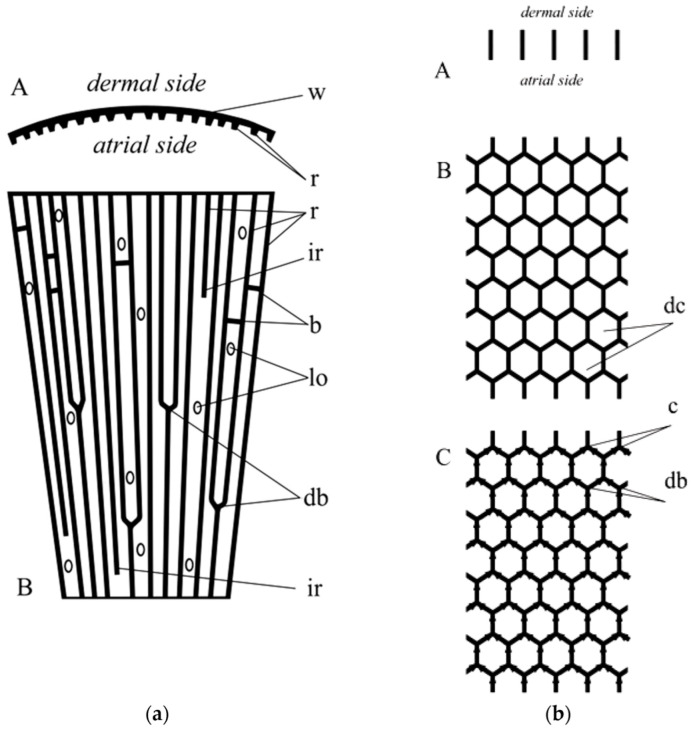
Figure 18.
(a) Scheme of the wall fragment of Lefroyella. A: horizontal section; w—wall; r—ridges. B: longitudinal section; r—ridges; ir—intercalary ridges; b—bridgees; lo—lateral oscula; db—dichotomous branching of ridges. (b) Scheme of the wall fragment of Aphrocallistes. A: horizontal section. B: longitudinal section; dc—honey-comb unit. C: longitudinal section, arrows show the suggested direction of the growth of the dictyonal skeleton; c—carina (line of fusion; db—dichotomous branching). It is of note here that dichotomous branching of actin filaments remains to be characteristic for this structural protein [67].

