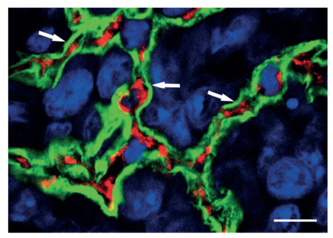
Figure 1.
As indication of vascular co-option is the preservation of alveolar architecture in the outer regions of lung metastases. The tumor mass is highlighted in this high-power confocal image of the HT1080 lung metastasis that has been labeled with podoplanin (green fluorescence), CD31 (red fluorescence), and TOTO-3 (blue fluorescence). Note the intact alveolar walls with normal layering (pneumocyte-capillary-pneumocyte) (arrows). Scale bar = 10 µm. (Reprinted with permission from Ref. [9]).

