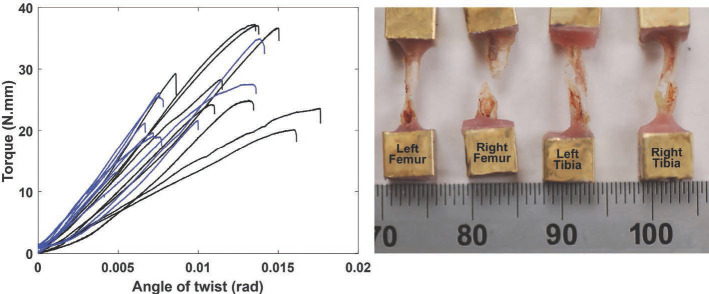
Figure 6.
Torque-angle of twist curves measured for irradiated (blue) and contralateral (black) femurs from the single dose (1×25 Gy) irradiation group (left). This composite plot illustrates the consistency in the slopes of the linear regions of the curves, and the general stiffening that was associated with radiation. The wide range in torsional stiffness and failure torque of the non-irradiated group was decreased with radiation. Composites from tibia tests and tests of the fractionated dosing groups were similarly clustered. Typical spiral fracture patterns resulting from the torsion test are shown for the femur and for the tibia (right). The ruler is showing units of millimeters.

